Structure and Function of a Cell and its Organelles
Living things consist of tiny living parts or compartments called cells. A cell itself consists of certain tiny parts or structures called organelles. These organelles perform various life functions within the cell.
This post will discuss the structure and function of the cell and its organelles.
Click here for the definition of the cell, the discovery of the cell, and cell theory
STRUCTURE AND FUNCTION OF A CELL
The cell provides structure and performs all the life functions of living things. Cells vary greatly in shape and structure.
Also, plant and animal cells are not alike. These may be disc-like, rectangular, flat, cuboidal, thread-like, branched, or even irregular. The structure of a cell is often related to the function it performs.
- Red blood cell in humans has the shape of a biconcave disc. This helps it to pass through narrow capillaries and transport oxygen.
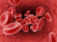
Red Blood Cells
- White blood cells in humans are of irregular shape, which is more like an amoeba. This helps it to squeeze out through capillary walls to fight pathogens.
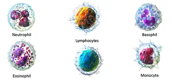
Various types of white blood cells
- A nerve cell is of an irregular shape with a long, thread-like structure. This helps it to conduct impulses from distant parts of the body to the brain.
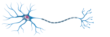
Nerve Cell
- The muscle cell is long and possesses elasticity. This feature helps it contract and relax to pull or squeeze the parts.
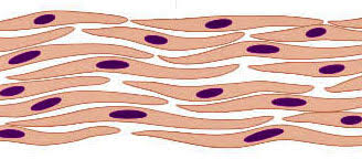
Muscle cells
- Guard cells of stomata in the leaves have a shape similar to that of kidney beans. This helps them to perform the function of opening and closing the pore.
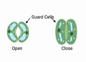
Stomata Guard Cell with open and closed pore
Back to >>>>Structure and Function of the Cell
GENERALIZED STRUCTURE OF CELL
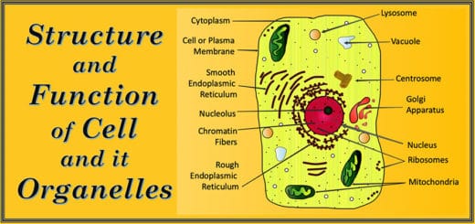
Various kinds of cells have different shapes and structures based on the functions they perform. Yet, all of them show some basic structural plans. This basic representation of cells, showing all features, is called a generalized cell. The generalized cell differs for plants and animals due to the presence or absence of certain parts or organelles.
Definition of Generalized Cell –
The generalized cell is the basic representation of cell showing all parts and organelles which can be present in any specialized cell. It is a hypothetical cell for a quick understanding of the basic structure and function of the cell and its organelles.
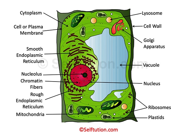
Structure of a Generalized Plant Cell showing various Organelles
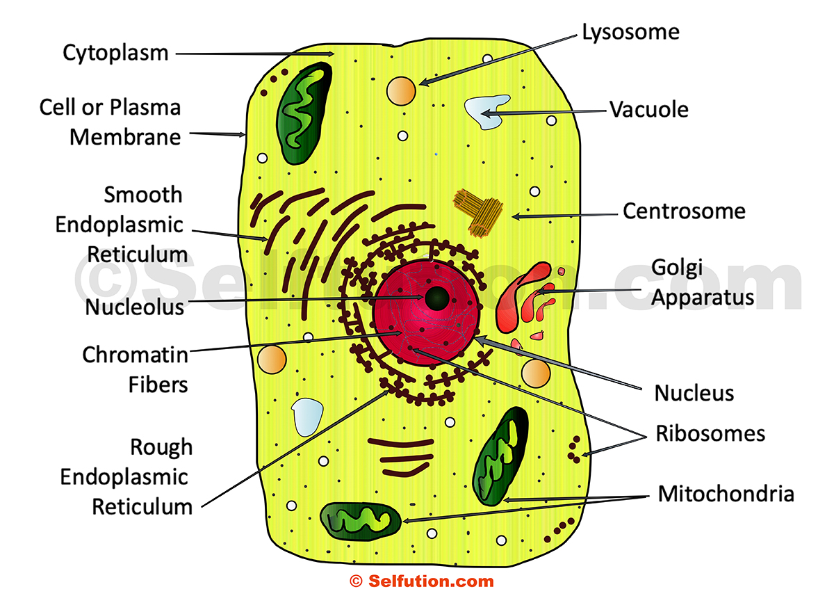
Structure of a Generalized Animal Cell with Various Organelles
All plant and animal cells consist of living and non-living parts.
The living part of the cell includes the cell membrane, the cytoplasm, and the nucleus. All three together are known as protoplasm. The non-living parts of the cell are granules and vacuoles.
The cytoplasm is embedded with tiny parts or structures called organelles. Organelle means ‘the little organs’. Organelles have definite structures and definite functions in the cell and have the same status in the cell as the organs have in the body of an animal or a plant.
Let us now discuss the different parts and organelles of the generalized cell of animals and plants in detail:
1. Cell Membrane or Plasma Membrane
The cell membrane is a very thin skin covering the cell. The cell membrane protects the cell and provides shape to it. It is made up of lipoprotein. There are very tiny holes in the cell or plasma membrane. It allows materials to enter and leave the cell through these tiny pores or openings. However, its permeability is selective. It means it allows certain substances to pass through it and prevents others.
The structure and function of a cell or plasma membrane are:
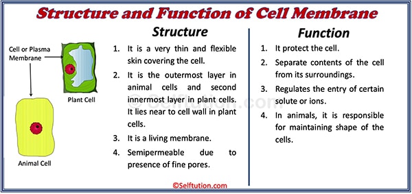
Structure and Function of the Cell or Plasma Membrane
Cell Wall: The cell wall is an extra covering that surrounds the cell membrane of a plant cell. It is made of stiff, non-living material called cellulose. The cell wall provides rigidity and protection to the cell. Unlike the cell membrane, it is freely permeable and allows all substances in solution form to pass through it. Animal cells do not have a cell wall.
The structure and function of a cell wall are:
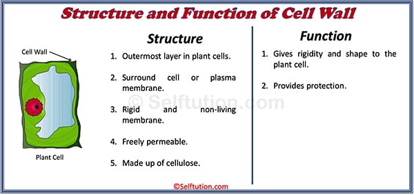
Structure and Function of the Cell Wall
2. Cytoplasm
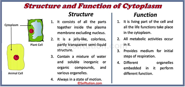
The cytoplasm is a jelly-like, semi-liquid structure occupying most of the inside of the cell. It occupies the space between the cell membrane and the nucleus. Under a microscope, it appears to be colorless, partly transparent, and somewhat watery. It is a living part of the cell, and all the life functions take place in the cytoplasm. The living cytoplasm is always in a state of motion. The cytoplasm contains many important tiny structures called organelles, which perform various life functions.
The structure and function of the cytoplasm are:
Structure and Function of Cytoplasm
3. Nucleus – The Control Center of the Cell
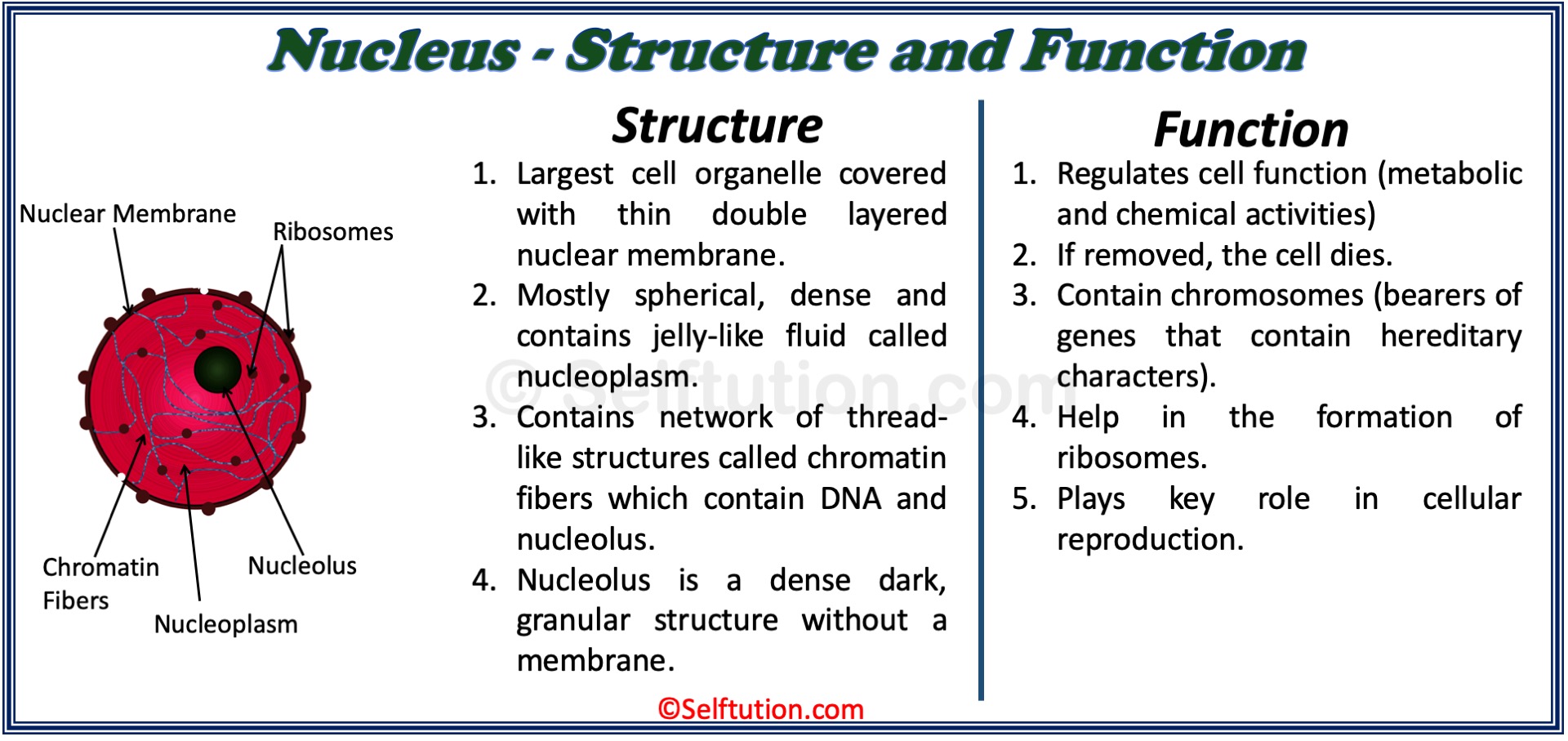
The nucleus is a spherical body present inside the cell. This structure is the control center of the cell, and its function is to regulate and coordinate the various life processes of the cell. Most cells have only one nucleus, but some cells, like those of muscles, have more than one nucleus.
The structure and function of the nucleus in the cell are:
Function and Structure of the Nucleus of the Cell
The nucleus comprises four parts – nuclear membrane, nuclear sap or nucleoplasm, nucleolus or nucleoli, and chromatin fibers.
Nuclear membrane – It is the delicate outermost covering layer of the nucleus. It separates the nucleus from the cytoplasm. A nuclear membrane, like the cell membrane, has tiny holes in it that allow the exchange of substances between the nucleus and the cytoplasm.
Nucleoplasm – It is the jelly-like fluid inside the nucleus. Chromatin fibers and nucleoli are embedded in the nucleoplasm.
Chromatin fibers – A network of thread-like structures called the chromatin network is present in the nucleoplasm. It consists of DNA (deoxyribonucleic acid) and proteins. At the time of cell division, the chromatin fibers develop into thick and ribbon-like, or rod-like structures called chromosomes. These chromosomes play an important role in carrying the genetic characters from the parents to the offspring.
Nucleolus or nucleoli – It is a dense, dark, granular structure without a membrane. It consists of RNA (ribonucleic acid) and proteins. It is the site of ribosome formation, thus, we can call it the factory of ribosomes.
Back to >>>>Structure and Function of the Cell
STRUCTURE AND FUNCTION OF CELL ORGANELLES
Organelle means ‘the little organs’. Organelles have definite structures and definite functions in the cell and have the same status in the cell as the organs have in the body of an animal or a plant.
a. Endoplasmic Reticulum
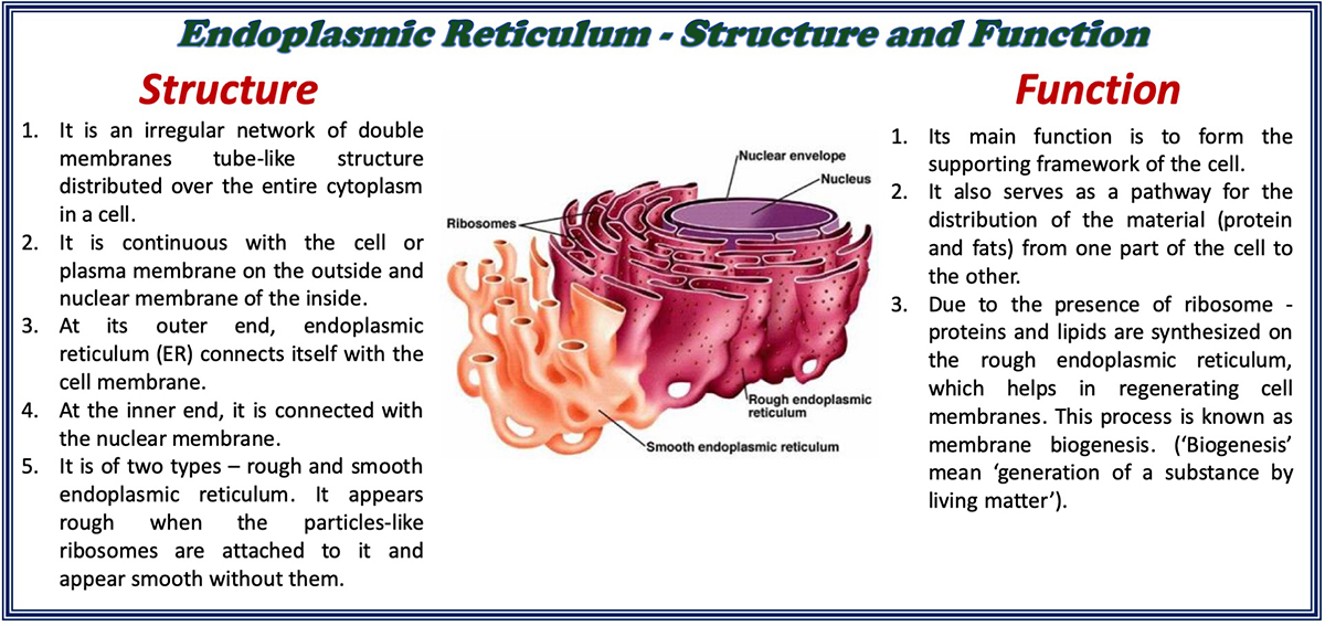
The endoplasmic reticulum (ER) is so fine in structure that we can view it only through an electron microscope. It is of two types – rough and smooth. The endoplasmic reticulum appears uneven due to the presence of particles like ribosomes attached to its surface. On the other hand, without them, it appears smooth. The function of the smooth and rough endoplasmic reticulum is to form the supporting framework of the cell. Due to the presence of ribosomes, the endoplasmic reticulum synthesizes proteins and lipids, which help in regenerating cell membranes. This process is known as membrane biogenesis. (‘Biogenesis’ means ‘generation of a substance by living matter).
The structure, characteristics, and function of the endoplasmic reticulum (ER) in the cell are:
Structure and function of the endoplasmic reticulum (ER)
b. Ribosomes
The ribosomes are numerous small granules either scattered freely in the cytoplasm or attached to the membranes of the endoplasmic reticulum. These are the single-walled dense, spherical bodies composed mainly of RNA. The most essential function of the ribosome is protein synthesis. Therefore, we also call them ‘factories’ or ‘sites’ for the synthesis of proteins.
c. Mitochondria
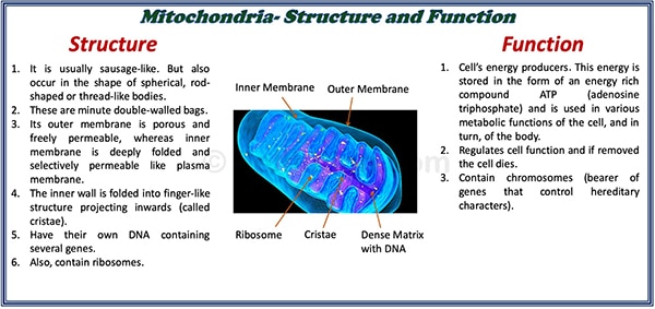
Mitochondria are the sites where cellular respiration occurs to release energy. Therefore, we can call mitochondria as “powerhouse of the cell or seat of cellular respiration”. The mitochondria are spherical or rod-shaped, or thread-like bodies. Mitochondria have ribosomes and DNA containing several genes. Due to this, mitochondria can even survive without a cell, thus it is a semi-autonomous organelles.
The structure and function of the mitochondria in the cell are:
Structure and Function of mitochondria in the cell
d. Golgi Apparatus
The Golgi apparatus acts as the delivery system of the cell. They occur in the form of granules, filaments, or rods that originate from the endoplasmic reticulum. These are tiny vesicles of different shapes arranged in parallel stacks (cisterns) located near the nucleus. The main function of the Golgi apparatus is the secretion of the cell, including enzymes, hormones, etc.
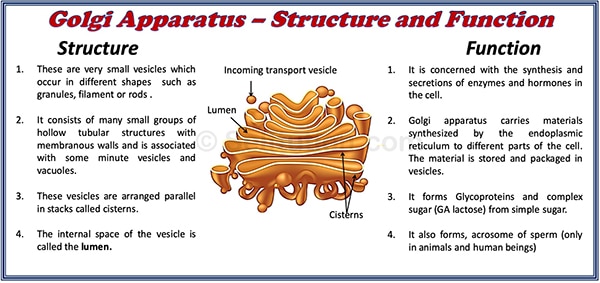
Structure and Function of the Golgi Apparatus in Cell
e. Lysosomes
Lysosomes are small vesicles of different shapes that bud off from the Golgi bodies. They contain 40 different types of enzymes. They help to keep the cell clean by digesting any foreign material as well as worn-out cell organelles. Lysosomes can do this because they contain powerful digestive enzymes (acid hydrolases) capable of breaking down all organic material. The enzymes are synthesized by the rough endoplasmic reticulum (RER). During the disturbance in cellular metabolism, for example, when the cell gets old or damaged, lysosomes may burst and enzymes are released to digest their cell. Hence, these are called “suicide bags”.
f. Centrosomes and Centrioles
A centrosome is present in animal cells only. It is located in a clear area of the cytoplasm close to the nucleus. Centrosomes consist of two barrel-shaped clusters of microfilaments, called “centrioles”. These two centrioles are arranged at right angles to each other. It develops spindle fibers during cell division, both in mitosis and meiosis. There are no centrosomes and centrioles in plant cells.
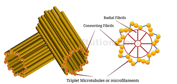
Structure of Centrosome and Centrioles
The structure of the Centrosome
- Centrosomes contain two centrioles arranged at right angles to each other.
- Centrioles are short, barrel-shaped bundles of microfilaments.
- Its microfilaments form a radiating star (aster) like structure during cell division.
The function of the Centrosome in the cell
- Initiate and regulate cell division.
- Forms spindle fibers, with the help of asters.
g. Plastids
Plastids are present only in plant cells. The function of plastids in the cell is to manufacture and store food in plants. They occur in different shapes – oval, spherical, and disc-shaped.
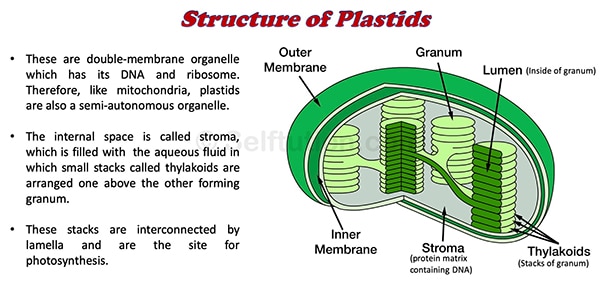
Structure of Plastids
Depending upon the color, the plastids are of three types – leucoplasts, chromoplasts, and chloroplasts.
- Leucoplasts are colorless plastids. They have no pigments. They store starch. The cells of potatoes have lots of leucoplasts in them.
- Chromoplasts are plastids of various colors – yellow, orange, and red. They are mostly present in the petals of flowers and fruits. The coloring substances (pigments) associated with them are xanthophyll (yellow) and carotene (orange-red).
- Chloroplasts impart a green color to a plant. They have green pigments called chlorophyll. Chloroplasts are abundant in parts exposed to light, for example, leaves. They also have other pigments such as orange and yellow. However, the chlorophyll present in large amounts masks these pigments. Their function is to trap solar energy and absorb carbon dioxide for the manufacture of starch and sugar during photosynthesis. Chloroplasts contain DNA and have the capacity to divide.
Back to >>>>Structure and Function of the Cell
NON-LIVING PARTS OF THE GENERALIZED CELL
a. Granules
Granules are small particles in the cytoplasm that store food particles, such as starch, glycogen, and fats.
b. Vacuoles
Vacuoles are clear spaces in the cytoplasm. They are filled with water and various substances in the solution state. A single membrane called ‘Tonoplast’ bound these bubble-like sacs. In plant cells, the vacuoles are usually quite large, and the liquid that they contain is called cell sap. The cell sap contains proteins, minerals, organic acids, etc. Vacuoles provide turgidity and rigidity to the cell. An animal cell does not have such prominent vacuoles, and vacuoles are fewer.
Back to >>>>Structure and Function of the Cell
For more such information, please visit our YouTube channel SELFTUTION
